Heart Diagram Class 10
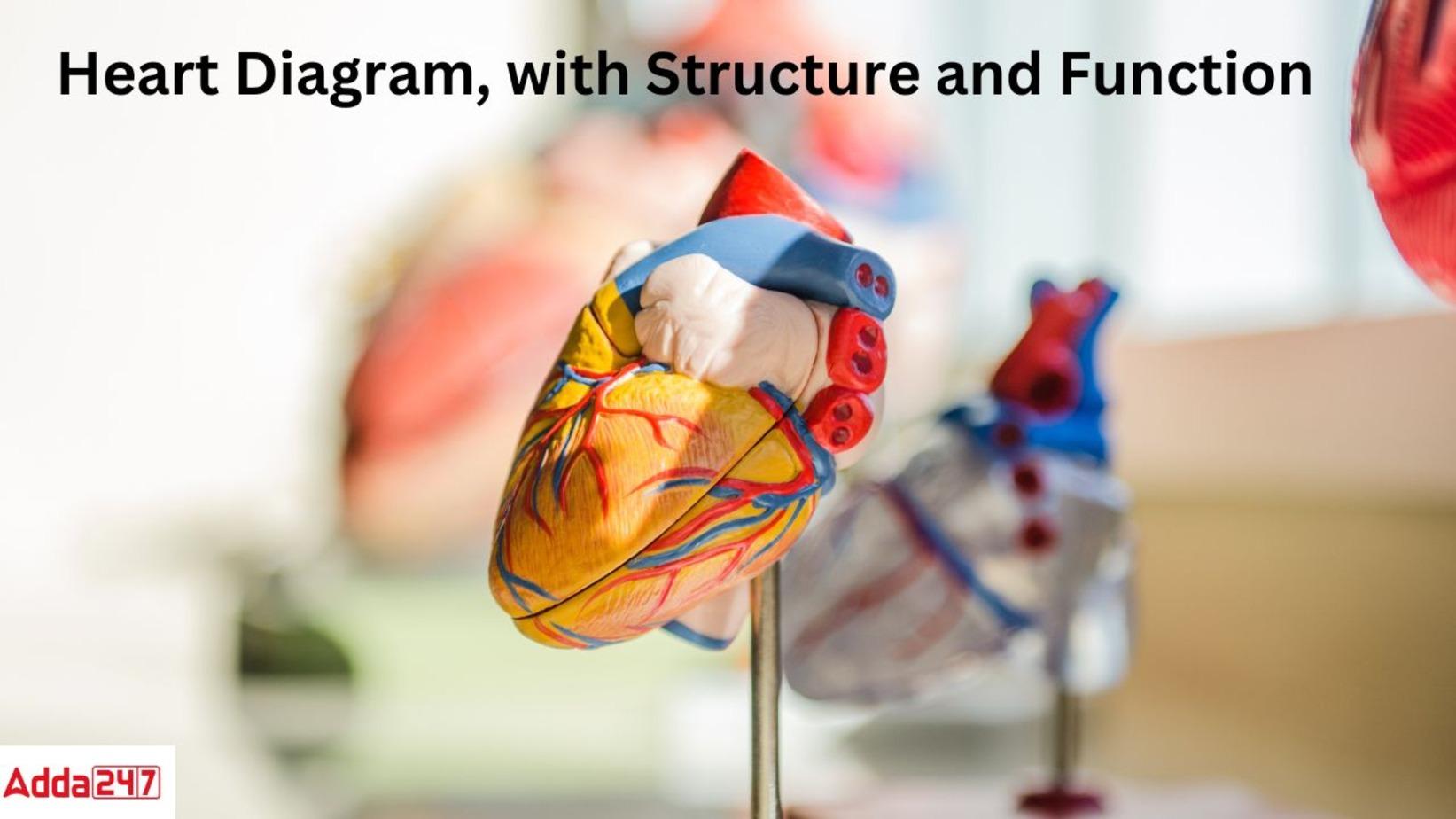
Drawing of Heart Diagram is one of the important and frequently asked questions in the Class 10 board exam. Also, the human heart diagram will help you to understand the heart structure and its blood circulation mechanism. Here we have provided the Simple Heart diagram labeled along with its structure and its blood circulation mechanism.
Easy Heart Diagram
Simple heart Diagram: The heart is one of the most vital organs in the human body. The heart pumps blood throughout the body. The blood that is pumped around the body supplies nutrients and oxygen, while also transporting metabolic waste products like carbon dioxide to the lungs. The human heart is situated in the center of the chest, between the lungs.
Human hearts have four chambers: lower left and right ventricles and upper left and right atria. The right atrium and ventricle are frequently referred to as the right heart, whereas their left counterparts are referred to as the left heart. The four valves that consist of the heart are the aortic valve, mitral valve, pulmonary valve, and tricuspid valve.
Easy Human Heart Diagram with labelling
Let’s understand the four chambers of the heart with a labeled Simple Heart Diagram.
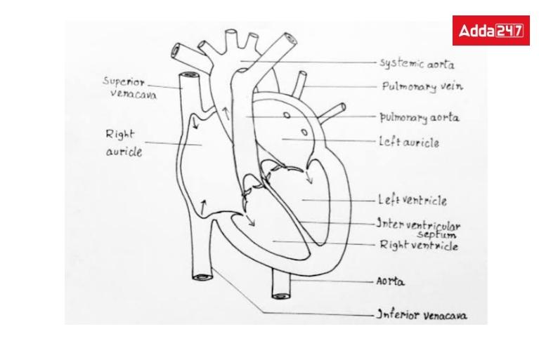
Human Heart Structure Diagram
The heart is the main and crucial organ of the human circulatory system. The human heart is conical. The human heart structure is complex due to its function. Let,s give a look at the heart structure.
- It is covered by the bilayer pericardium. It is located in the middle of the lungs of the thoracic cavity in the case of the curtain. The wall is made up of three layers: the epicardium, which acts as a protective outside layer, the myocardium, which is a muscular middle layer, and the endocardium, which is a thin inner layer.
- The human heart consists of four chambers. The upper two chambers are the left and right atrium and the lower two chambers are the left and right ventricles. The atria are separated by two inter-atrial septa and the ventricles are separated by two inter-atrial septa.
- The right atrium is connected with one superior and inferior vena cava and the left atrium with four pulmonary veins. The left ventricle gives off the aorta and the right ventricle gives off the pulmonary artery.
- At the junction of the right atrium and the right ventricle is the tricuspid valve and at the junction of the left atrium and the left ventricle is the bicuspid valve or the mitral valve.
- Similarly, the junction of the right ventricle and the pulmonary artery has the pulmonary valve and the junction of the left ventricle and the aorta has the aortic valve. The latter valves are called semilunar valves as they are crescent-shaped.
Diagram of Heart
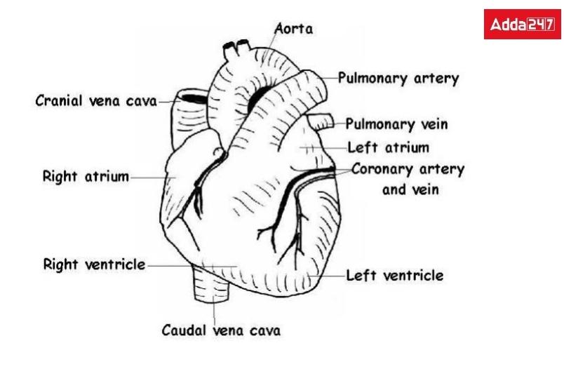
Human Heart Diagram labeled
To understand Heart structure more clearly. Let’s give a look at the heart diagram.
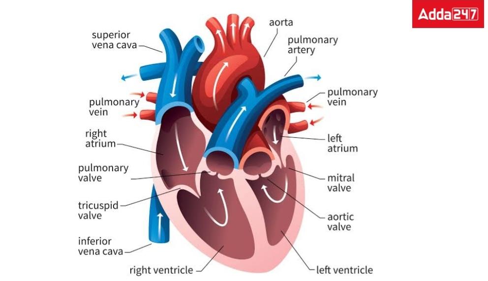
Human Heart Functions
Heart Function: Human heart plays many vital roles in the body for life-sustaining. The main function of the heart is to ensure a steady flow of blood throughout the body. As a result, the cells and tissues receive fresh oxygen and nutrients. The heart performs the following primary functions:
- Blood circulation throughout the body is one of the heart’s primary functions.
- The human heart is one of the many organs in the body that receives oxygen, hormones, glucose, and other substances through the blood.
- The heart also makes sure that the body’s blood pressure is kept at a healthy level.
- It plays a vital role in maintaining a stable heart rate for healthy living.An adult’s heart beats an average of 72 times per minute.
Human Heart Structure and Function- Mechanism of blood circulation through Heart
Due to the complex heart structure and function, the Blood circulation system in the heart includes several stages as follows
Mechanism of blood circulation through the heart
- The contraction of the heart is called systole and expansion is called diastole. The heart circulates blood by repeatedly contracting and expanding in a certain rhythm.
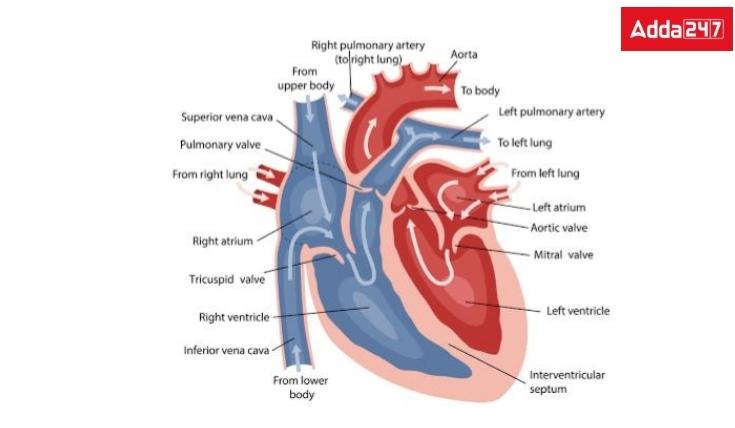
- When the atria of the heart are in diastole, contaminated blood (carbon dioxide colored) from the whole body enters the right atrium through the superior and inferior vena cava, and pure blood (oxygenated blood) from the lungs enters the left atrium through the pulmonary veins.
- When the atria are filled with blood, the atrial systole occurs, so that the contaminated blood from the right atrium enters the right ventricle and the pure blood from the left atrium enters the left ventricle, while the mitral and tricuspid valves are open.
- When the ventricles are full of blood, then systole occurs. Then the bipartite and tripartite lids close and the semilunar lids open.
- As a result, contaminated blood from the right ventricle goes to the lungs through the pulmonary artery and the pure blood from the left ventricle enters the aorta and spreads throughout the body through various arteries.
- And this whole process is repeated again and again to maintain steady and oxygenated blood flow throughout the body.
Related Posts
| What is cell | |
| Simple Microscope | Respiration- In Plants and Human Beings, Equation |
| Vegetative propagation | Paramecium Diagram ,classification |
Human Heart Diagram-FAQs
Q.What is the function of the heart?
The major organ of the human blood circulatory system is the heart. The main function of the heart is to ensure a steady flow of blood throughout the body. As a result, the cells and tissues receive fresh oxygen and nutrients It maintains blood pressure, regulates heart rate, and pumps blood throughout the body.



 How to Check CUET UG 2025 Result in 1 Mi...
 Documents Required for NEET Counselling ...
 ACPDC Merit List 2025 Out @acpdc.gujarat...

